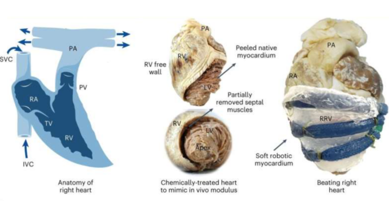
Engineers from the Massachusetts Institute of Technology (MIT) have built a robotic replica of the heart’s right ventricle, which they hope will help study heart disorders and test therapeutic interventions.
The robo-ventricle merges actual heart tissue with synthetic, balloon-like artificial muscles. This function was introduced to offer scientists precise control over contractions and the ability to observe the intricate functioning of natural valves.
The artificial ventricle, known as RRV, not only replicates healthy states but can also be adjusted to simulate conditions associated with right ventricular dysfunction, such as pulmonary hypertension and myocardial infarction. The team employed the model to evaluate cardiac devices, showcasing its potential for testing and refining treatment strategies.
Read more: Irish hospital completes first robot-guided heart surgery in UK and Ireland
Manisha Singh, a postdoc at MIT’s Institute for Medical Engineering and Science (IMES), said:”The right ventricle is particularly susceptible to dysfunction in intensive care unit settings, especially in patients on mechanical ventilation.
“The RRV simulator can be used in the future to study the effects of mechanical ventilation on the right ventricle and to develop strategies to prevent right heart failure in these vulnerable patients.”
The right ventricle, often described as a “ballerina” due to its thinner muscle and complex motion, has proven challenging to assess accurately using conventional tools. This model, designed to replicate the ventricle’s anatomical intricacies and pumping function, addresses the limitations of existing diagnostic methods.
The team’s approach involved incorporating real heart tissue into the model, preserving its delicate structures that are difficult to reproduce synthetically.
By explanting a pig’s right ventricle and encasing it in a silicone wrapping with embedded balloon-like tubes, the researchers achieved a realistic simulation of the ventricle’s contractions and internal structures.
RRV’s pumping ability, which was designed to be akin to that observed in live, healthy animals, was demonstrated by infusing it with a blood-like liquid. The researchers could also manipulate the model to mimic various cardiac conditions, such as irregular heartbeats and hypertension, providing a versatile platform for experimentation.
The robo-ventricle’s potential extends to testing cardiac devices, MIT has said, with the team demonstrating the successful implantation of ring-like medical devices to repair the tricuspid valve.
By simulating conditions like leaky valves, the researchers observed how different devices improved fluid flow, showcasing the model’s utility in training surgeons and interventional cardiologists.
While the current version of the RRV can simulate realistic functions during the course of a few months, ongoing efforts are focused on extending its performance for continuous use.
The researchers are collaborating with device designers to test prototypes on the artificial ventricle, potentially expediting their journey to clinical applications.
Looking ahead, there are plans to pair the RRV with a similar artificial model of the left ventricle, with the intention of manufacturing a fully tunable artificial heart that they hope could have major implications for medical science.


